01
May 2026
ARVO 2026: Clinical OCX Imaging
ARVO POSTER 0201: Dual optical coherence tomography and microscopy for in vivo assessment of corneal pathologic changes
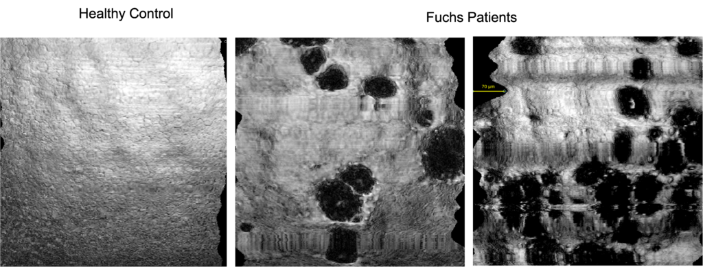
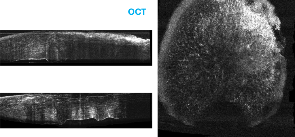
We evaluated the performance of a dual-modality imaging system (OCX) integrating optical coherence tomography (OCT) and Optical Coherence Microscopy (OCM) for in vivo assessment of corneal pathologic changes. OCM enabled high-resolution en-face visualization of microstructural features, including endothelial cells and guttate in FECD (Fuchs Endothelial Corneal Dystrophy) subjects. OCT volume provided wide-field corneal architecture.
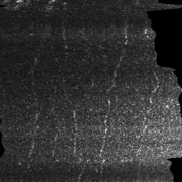
Endothelium imaged in vivo with OCX® (OCM mode)
OCX® OCT of corneal infections: Fungal: Fusarium spp
OCX® OCM imaging of corneal infection: Fungal: Fusarium spp
OCX® imaging of Ocular Surface Squamous Neoplasia (OSSN)
OCX® imaging of nerves in Dry Eye